Why Cell Culture Protocols Might Be Producing the CPE They’re Looking For

For decades, virologists have relied on a simple visual signal in cell culture to tell them a virus is present: cytopathic effect, or CPE. The cells round up, become refractile, form syncytia, detach, or show vacuoles and granularity. It’s been the workhorse endpoint since the 1950s. But what if a key part of the standard protocol itself helps create the very morphology being scored as “positive”?
I’ve spent time reviewing the institutional guidelines that shape how virus isolation is done worldwide. What emerges is a consistent structural pattern in the protocols themselves—and a subtle but important feedback loop in how those protocols have evolved.
What the institutional guidelines actually say
(sorry for the crappy table, substack doesn’t have table feature).
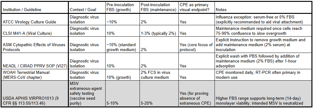
Every major body that sets the rules for diagnostic and research virus isolation uses essentially the same playbook. Cells are grown to near-confluence in “growth medium” (typically 8–10% fetal bovine serum). Then, at or immediately after inoculation with the clinical specimen or viral stock, the medium is switched to “maintenance medium” containing far less serum.
However, there is a curious outlier, highlighted in grey. Notice how in this case instead of FBS reduction at inoculation, the FBS could be increased. What mental model can we use to understand why the protocols would diverge in terms of the change of FBS concentration between growth and maintenance medium?
First we need to understand how the outlier might be different. MVS extraneous-agent safety testing produces a ‘positive’ results when no CPE is present in the culture. This contrasts the virus isolation protocols which produce a ‘positive’ result when CPS is present in the culture.
The data suggest a straightforward, testable model:
FBS concentration in the maintenance phase is a major variable that influences whether CPE-like morphology appears.
High serum (10% or more) keeps cells healthier and more stable → fewer non-specific changes.
Low serum (2% or less) stresses the monolayer → increased rounding, refractility, detachment, vacuoles, granularity.
This model predicts exactly the protocol divergence we observe: isolation methods drift toward lower serum (making the desired “positive” easier to see), while purity/safety assays tolerate higher serum (making the desired “negative” easier to achieve).
It also explains why the pattern has persisted for seventy years. Once CPE was accepted as the visual hallmark of viral infection in the 1950s, every subsequent protocol is built on that foundation. Conditions that reliably produced the expected morphology became standardized. Over time the system selected for itself.
Does the practice align with the protocol?
One obvious question: does the laboratory practice match the protocol?
I have spent considerable time looking for papers that actually list or allude to both the pre and post inoculation FBS concentration. Here is a list of those papers. Not all follow the virus isolation protocol, but the general trend is that the protocol is followed.
Isolation and characterization of a bat SARS-like coronavirus that uses the ACE2 receptor
Severe Acute Respiratory Syndrome Coronavirus 2 from Patient with Coronavirus Disease, United States
A Vero-cell-adapted vaccine donor strain of influenza A virus generated by serial passages
Enhanced Isolation and Detection of COVID-19 in Hospitalized Patients Undergoing Antiviral Therapy
Antimycotic-Antibiotic Amphotericin B Promotes Influenza Virus Replication in Cell Culture
Cell Culture Extraction and Purification of Rabies Virus Nucleoprotein
Isolation and Molecular Characterization of Feline Herpesvirus 1 from Naturally Infected Korean Cats
Isolation and genetic characterization of MERS-CoV from dromedary camels in the United Arab Emirates
Breastfeeding by chikungunya virus-infected dams confers resistance to challenge in the offspring
Trypsin enhances SARS-CoV-2 infection by facilitating viral entry
Characterization of a small plaque variant of West Nile virus isolated in New York in 2000
Mechanisms of establishment of persistent SARS-CoV-infected cells
What this means for virology
CPE was the original, foundational observable used to define and characterize viruses. Every subsequent layer of virology—electron microscopy, genomic sequencing, immunological assays, antigen detection—rested on the assumption that the CPE being observed was specifically caused by the virus.
If a significant portion of that CPE is instead a non-specific response to the culture conditions (particularly the routine drop to low FBS), then the entire chain of evidence becomes questionable.
Viral genome databases may contain sequences amplified from cultures whose “infection” was partly or wholly a serum-starvation artifact.
Immunological reagents developed against “viral” proteins may in fact be detecting stress-induced cellular components.
Electron micrographs labeled as “virions” may include exosomes or other stress particles released under low-serum conditions.
This means that the primary visual assay used for decades to detect and study them has a built-in confounder that was never fully characterized.
The protocols are the product of decades of practical, incremental refinement under real scientific and institutional incentives. But those incentives have shaped the tools in ways that deserve careful scrutiny.
The question is no longer whether CPE is virus-specific. The question is how much of what we have called “viral CPE” has always been, at least in part, a predictable response to the very conditions we created to look for it.
"..If a significant portion of that CPE is instead a non-specific response to the culture conditions (particularly the routine drop to low FBS), then the entire chain of evidence becomes questionable..."
That`s the gold! "Isolation" of viruses has many critics, some of which are mad but some have valid arguments....